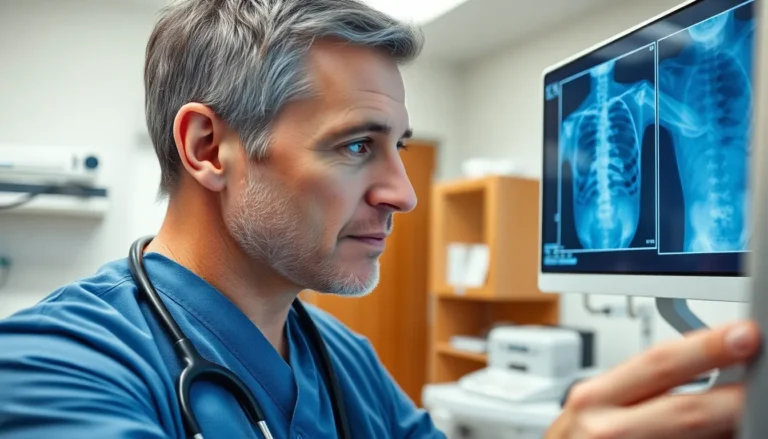
Understanding the x-ray stages of bone healing is crucial for both medical professionals and patients alike. When a bone fractures, the body embarks on a remarkable journey to repair itself, and x-rays play a vital role in monitoring this process. From the initial inflammatory phase to the final remodeling stage, each phase reveals unique changes that can be tracked through imaging.
These stages not only help in assessing the healing progress but also guide treatment decisions. By recognizing the signs of healing on x-ray, healthcare providers can ensure patients receive the best care possible. This article will delve into the intricate stages of bone healing as seen through x-ray imaging, shedding light on how these images inform recovery and contribute to successful outcomes.
Understanding Bone Healing
Bone healing occurs in distinct stages, each characterized by specific physiological processes. Medical professionals utilize x-ray imaging to visualize these stages and assess healing progress.
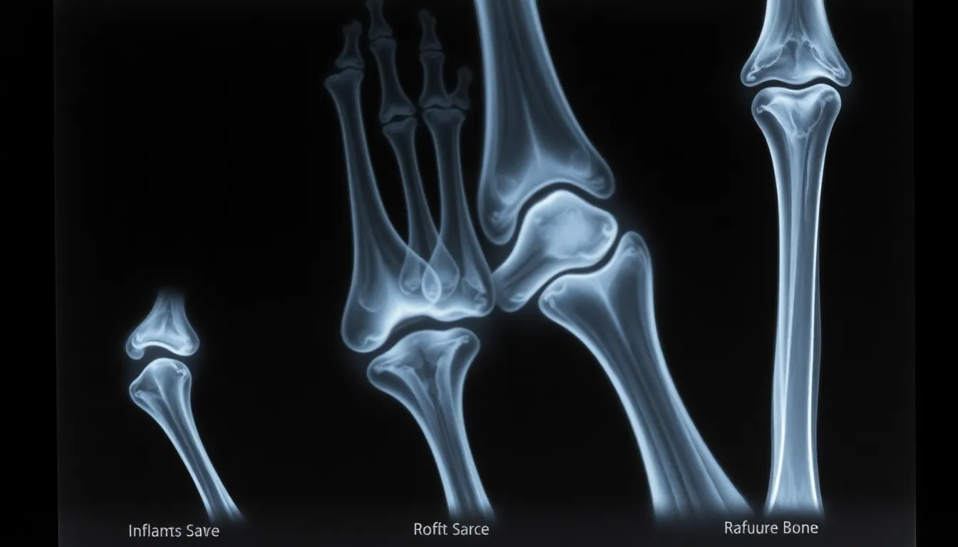
- Inflammation Stage: This initial phase begins immediately after a fracture. It involves the formation of a hematoma, which serves as a scaffold for subsequent healing. X-rays during this stage may show a clear fracture line, indicating the separation of bone fragments.
- Soft Callus Formation: Within a week, the body begins forming a soft callus made primarily of collagen and cartilage. During this phase, x-rays may still reveal the fracture line but show new tissue development around the break.
- Hard Callus Formation: This stage can last from several weeks to a few months. The soft callus transforms into a hard callus as bone tissue replaces the cartilage. X-rays display increased radiopacity, indicating the presence of new bone formation bridging the fracture gap.
- Remodeling Stage: The final phase can take several months to years. The hard callus becomes remodeled into the bone’s original shape and structure. X-rays reflect this stage through the gradual disappearance of the callus and a return to normal bone contour.
Understanding these stages enhances the ability to evaluate healing accurately and adjust treatment plans as necessary, ultimately improving patient outcomes.
Xray Stages of Bone Healing
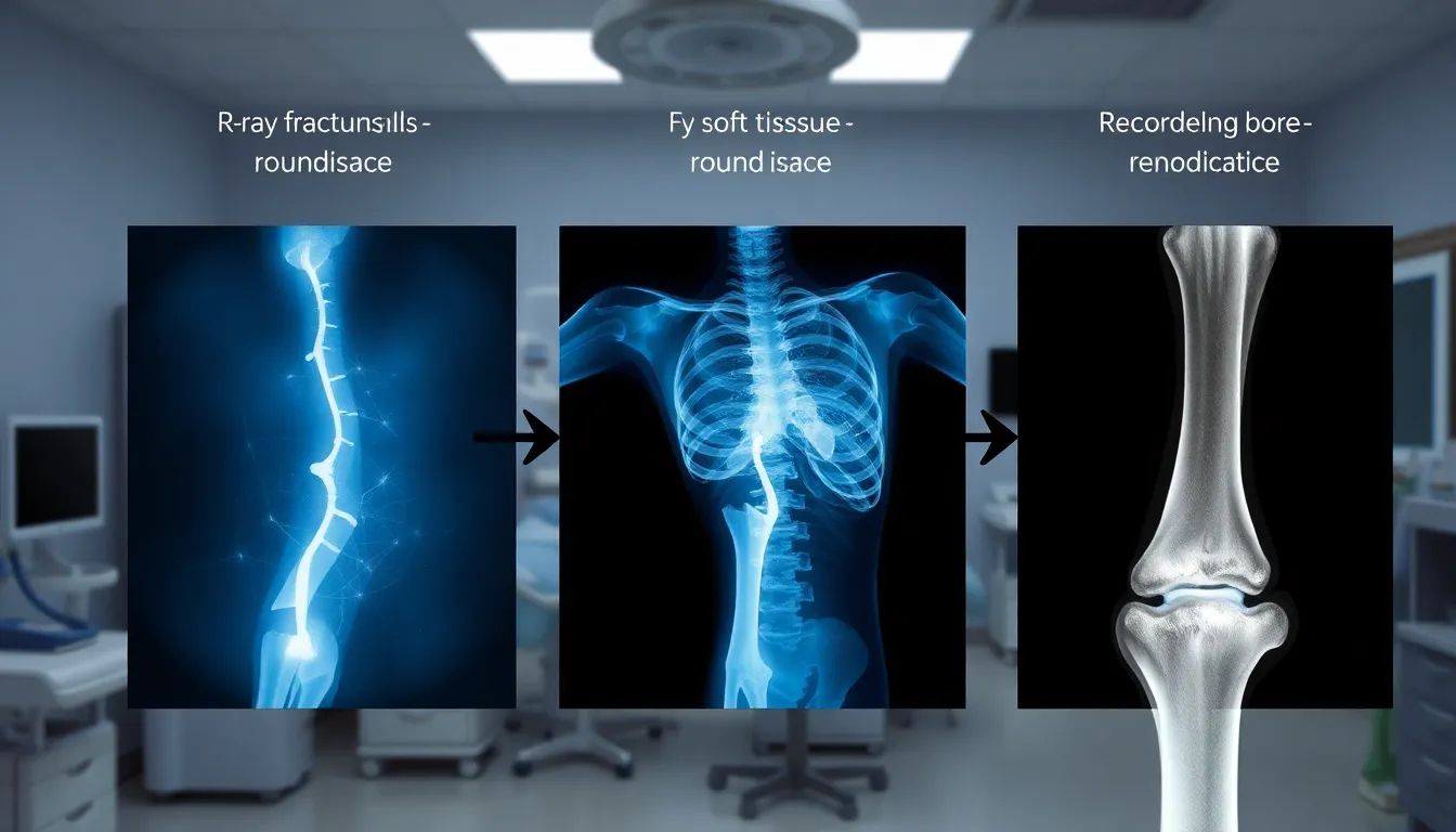
X-rays provide critical insights into the progression of bone healing, capturing distinct changes in each stage. Understanding these stages aids in assessing the healing process and adjusting treatment when necessary.
Inflammatory Stage
The inflammatory stage begins immediately after a fracture occurs. Hematoma formation acts as a temporary scaffold, initiating the healing process. X-rays taken during this stage often show a clear fracture line, reflecting the separation of bone fragments and the body’s immediate response to injury.
Repaired Stage
The repaired stage, often referred to as soft callus formation, typically starts within one week post-fracture. The body produces new tissue primarily composed of collagen and cartilage. X-rays during this phase continue to reveal the fracture line, but they also highlight the development of new soft tissue surrounding the site, demonstrating the body’s efforts to stabilize the fracture.
Remodeling Stage
The remodeling stage can extend from several months to years, as the hard callus undergoes gradual transformation back to the original bone structure. This phase involves the replacement of the soft callus with mature bone tissue. X-rays will show increased radiopacity that signifies new bone formation bridging the fracture gap. Over time, X-rays illustrate the gradual disappearance of the callus and a return to normal bone contour, indicating successful remodeling.
Importance of Xrays in Bone Healing
X-rays play a crucial role in monitoring the bone healing process, providing insights throughout the recovery stages. X-ray imaging allows medical professionals to assess the effectiveness of treatments and make necessary adjustments to optimize patient outcomes.
- Real-Time Monitoring: X-rays provide real-time visualization of the healing stages. They enable an accurate assessment of fracture alignment and the formation of callus tissue, ensuring that the healing process proceeds smoothly.
- Phase Confirmation: X-rays confirm the transition between stages of healing. They highlight the development from hematoma to soft callus and, eventually, to hard callus, allowing timely interventions if any complications arise.
- Assessment of Complications: X-ray imaging helps identify potential complications such as malunion or nonunion of fractures. Early detection of issues through X-rays facilitates prompt therapeutic actions to correct or enhance the healing process.
- Guidance for Surgical Decisions: X-rays assist surgeons in determining the need for surgical intervention. Clear imaging of the healing stages informs decisions about fixation devices, grafting, or other surgical options based on the healing progress observed.
- Documentation and Progress Tracking: X-ray images serve as essential records for tracking the recovery journey. They provide visual documentation of healing milestones, supporting both clinical assessments and patient education on their recovery progress.
X-rays are integral to understanding and managing bone healing, ultimately enhancing recovery outcomes through informed decision-making.
Factors Affecting Bone Healing
Recovery from a fracture depends on various factors. These factors can significantly influence the efficiency and effectiveness of the healing process.
Age
Age impacts bone healing. Younger individuals often experience faster healing due to increased cellular activity and higher metabolic rates. Older individuals, on the other hand, tend to have slower healing processes because of decreased bone density and regenerative capabilities.
Nutrition
Nutrition plays a critical role in bone health. A diet rich in calcium and vitamin D supports optimal healing. Calcium strengthens bones, while vitamin D enhances calcium absorption. Deficiencies in these nutrients can delay healing.
Blood Supply
Blood supply directly affects healing. Adequate blood perfusion brings essential nutrients and oxygen to the fracture site. Impaired circulation, such as in cases of vascular disease or external compression, can hinder the healing process.
Overall Health
Overall health status contributes to healing outcomes. Chronic conditions like diabetes or autoimmune disorders can impair the body’s healing capacity. Obesity can also complicate healing due to increased mechanical stress on bones.
Smoking and Alcohol Consumption
Smoking negatively impacts bone health. It decreases blood flow and reduces the body’s ability to deliver vital nutrients to the bone. High alcohol consumption can also interfere with bone repair by inhibiting calcium absorption and disrupting hormone levels.
Activity Level
Activity level influences healing. Gentle movement can promote circulation and encourage healing, while excessive stress on a fracture can lead to complications. Proper rehabilitation and adherence to prescribed activity levels support recovery.
Medications
Certain medications can affect bone healing. Non-steroidal anti-inflammatory drugs (NSAIDs) may interfere with inflammation, which is crucial for the healing process. Corticosteroids can weaken bone tissue and prolong recovery.
Fracture Characteristics
Fracture characteristics, including type and location, impact healing. Complex fractures may require longer healing times and more intensive treatment. In contrast, simple fractures typically heal faster.
These factors interact with the healing stages, underscoring the importance of a multidisciplinary approach to fracture management and recovery.